Controlling Brain Cells with Light: How Pond Algae Inspired a Nobel Prize in Medicine
The Nobel Assembly at Karolinska Institutet awarded the 2026 Nobel Prize in Physiology or Medicine to bioengineer and psychiatrist Karl Deisseroth, biophysicist Peter Hegemann, and molecular biologist Georg Nagel for their pioneering discoveries concerning light-gated ion channels and the development of optogenetics. Their work transformed modern neuroscience by establishing a method to manipulate specific nerve cells using targeted beams of light, solving a fundamental challenge in understanding the human central nervous system.
The breakthrough bridges two distant corners of biology: the light-tracking machinery of single-celled freshwater algae and the millisecond-scale electrical circuitry of the mammalian brain. By isolating the algal protein channelrhodopsin and transplanting it into neurons, the laureates engineered a biological switch that enables researchers to activate or silence individual neural circuits with cellular specificity and temporal precision.
The Laureates and the Path to the Nobel Prize
The scientific journey recognized by the Nobel Committee spans more than three decades, beginning with basic curiosity-driven microbiology in Germany and culminating in advanced bioengineering in the United States.
Peter Hegemann, working at the Max Planck Institute for Biochemistry and later at Humboldt University of Berlin, investigated phototaxis—the navigational movement of microorganisms toward or away from light. Partnering with Georg Nagel, then at the Max Planck Institute of Biophysics and subsequently the University of Würzburg, Hegemann sought the molecular basis behind the rapid light reactions of green algae. Their joint experiments demonstrated that an algal photoreceptor protein could directly channel electrical currents across cellular membranes without requiring intermediate signaling enzymes.
At Stanford University, Karl Deisseroth, an active clinical psychiatrist and bioengineer, recognized the clinical bottleneck in brain research. Traditional psychiatric and neurological interventions—ranging from pharmaceutical medications to electroconvulsive therapy and deep-brain electrical stimulation—affected broad brain areas indiscriminately, causing widespread off-target effects. Collaborating with Hegemann and Nagel, Deisseroth’s laboratory successfully introduced the genetic blueprints of channelrhodopsin into mammalian neurons, proving that pulses of blue light could reliably fire action potentials inside intact, living brain tissue.
The resulting discipline, coined “optogenetics,” revolutionized investigative neurology. Laboratories worldwide rapidly adopted the technology to map complex behavior, delineate motor control pathways, study neuropsychiatric conditions, and explore memory formation.
The Biology of Light Perception in Pond Algae
All living organisms rely on environmental cues to survive. For photosynthetic life, solar radiation is an energetic imperative. Yet, long before multicellular animals evolved compound eyes or vertebrate retinas, single-celled micro-organisms perfected self-contained sensory systems capable of processing ambient illumination.
The single-celled green alga Chlamydomonas reinhardtii swims through freshwater environments via two whip-like flagella. To survive, it must navigate toward optimal light zones for photosynthesis while steering clear of intense ultraviolet and solar radiation that could generate toxic reactive oxygen species and damage its photosynthetic machinery.
Chlamydomonas accomplishes this balancing act using an organelle called the eyespot apparatus. Unlike mammalian vision, which requires complex retinas, optic nerves, and dedicated visual cortices to form images, the algal eyespot acts as a directional light antenna. When light strikes the eyespot, the alga alters the beating pattern of its two flagella within milliseconds, turning its body toward or away from the light source.
Biologists previously presumed that microalgae perceived light using complex signaling cascades analogous to the visual rhodopsins found in human photoreceptor rod and cone cells. In the human eye, light absorption triggers an enzymatic biochemical cascade that indirectly closes ion channels. In the 1990s and early 2000s, Hegemann and Nagel proved that Chlamydomonas took an entirely different evolutionary route: its sensor and its electrical gate were housed inside a single molecular machine.
Channelrhodopsin: A Self-Contained Optogenetic Gate
The core protein discovered by Hegemann and Nagel is channelrhodopsin. Built directly into the outer plasma membrane of the algal cell, channelrhodopsin spans the lipid bilayer seven times, forming a serpentine transmembrane structural motif known as a seven-transmembrane helix bundle.
A protein channel alone, however, cannot detect visible light. Channelrhodopsin requires a light-absorbing chemical cofactor: a vitamin A derivative known as retinaldehyde (or retinal). Retinaldehyde nestles deep within the protein pocket of channelrhodopsin, covalently linked to a specific lysine residue via a protonated Schiff base.
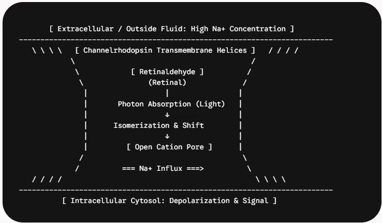
When a photon of appropriate wavelength (predominantly blue light around 470 nanometers) strikes the retinaldehyde molecule, the cofactor absorbs the energy and undergoes an immediate chemical shape change called photoisomerization.
The physical twisting of retinaldehyde exerts mechanical tension on the surrounding amino acid helices of channelrhodopsin. This structural shift forces open a narrow aqueous pore through the center of the protein. Positively charged ions—principally sodium ($Na^+$), protons ($H^+$), and calcium ($Ca^{2+}$)—instantly rush through this opening, moving along their electrochemical gradient from the outside water into the cell.
In Chlamydomonas, this sudden influx of positive ions alters the electrical charge across the plasma membrane, generating a photoreceptor current that commands the flagella to pivot and steer the cell.
Neurobiology Fundamentals: Why Ions Matter to the Brain
To understand why an algal ion gate earned the highest honor in physiology and medicine, one must examine how the 86 billion neurons in the human brain communicate.
Neurons are electrically excitable cells. Under resting conditions, specialized metabolic pumps actively export positive sodium ions out of the neuron while retaining potassium ions inside. This distribution creates an electrical charge imbalance across the neural membrane:
-
The interior of a resting neuron maintains a negative charge relative to the extracellular environment, typically hovering at approximately -70 millivolts (mV).
-
When a neuron receives stimulating neurotransmitters from upstream neighbors, specialized gates crack open, allowing sodium ions to flow inside.
-
If enough positive charge enters to raise the internal voltage past a critical threshold (typically around -55 mV), voltage-gated ion channels fly open down the axon.
-
The neuron discharges a rapid electrical spike known as an action potential, releasing chemical neurotransmitters at its synaptic terminals to signal the next neuron in the circuit.
For decades, neuroscientists struggled to understand how these action potentials translate into thought, emotion, sensory perception, and motor action. The fundamental experimental hurdle was specificity.
If researchers stimulated the brain using metal microelectrodes, the electrical current spread non-selectively, firing every neuron, passing axon, and neighboring cell within reach. If researchers introduced neuroactive drugs, the chemical washed across vast brain regions slowly and lingered for minutes or hours, far too coarse to match the millisecond operational speed of neural calculations.
The Breakthrough: From Pond Water to Mammalian Brains
The convergence between microbial biophysics and brain engineering occurred when Hegemann, Nagel, and Karl Deisseroth asked a bold question: What happens if you take the gene encoding channelrhodopsin from Chlamydomonas and place it inside a mammalian nerve cell?
Because retinaldehyde is naturally abundant in mammalian brain tissue in the form of vitamin A, researchers did not need to feed foreign chemical cofactors to laboratory animals. They only needed to deliver the genetic code of the channelrhodopsin protein.
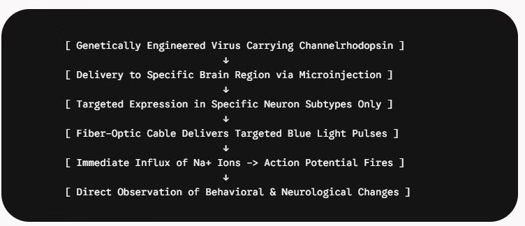
Using engineered, harmless viral delivery vectors—such as adeno-associated viruses (AAV)—Deisseroth’s team packaged the algal DNA alongside specialized genetic promoter sequences. These promoters acted like postal delivery addresses, ensuring that only targeted subtypes of neurons (such as dopamine-producing neurons, inhibitory GABAergic interneurons, or excitatory glutamatergic projection cells) translated the gene into physical proteins.
Once delivered, the neuron’s cellular machinery manufactured channelrhodopsin and slotted it into its own outer membrane. When researchers shined a blue light pulse delivered via hair-thin fiber-optic threads implanted in the brain, the algal channels snapped open. Sodium ions poured into the mammalian neuron, driving its membrane voltage above the activation threshold within milliseconds.
When the light switched off, the channels closed, returning the neuron to its resting state. For the first time in medical history, scientists possessed a remote control to command specific neural pathways with millisecond precision.
Optogenetics vs. Classical Neuroscience Methodologies
To appreciate the scale of this breakthrough, consider how optogenetics compares directly to the tools neuroscientists relied upon throughout the 20th century:
| Scientific Parameter | Electrical Microstimulation | Pharmacological Injections | Surgical Lesions / Ablations | Optogenetic Control |
| Primary Mechanism | External electrical field depolarization | Receptor agonist/antagonist binding | Physical or chemical tissue destruction | Light-gated algal ion channel activation |
| Cell-Type Specificity | None; activates all surrounding cell bodies and fibers | Moderate; targets receptors, but receptors exist across many cell classes | Zero; destroys all local cell types and passing tracts | Extreme; restricted via cell-specific genetic promoters |
| Temporal Precision | Milliseconds | Minutes to hours | Irreversible / Permanent | Milliseconds; synchronized to optical laser pulses |
| Spatial Precision | Coarse; current spreads through conductive tissue | Diffuse; subject to chemical diffusion gradients | Regional; limited by surgical cut or chemical spill | Microscopic; restricted to illuminated, opsin-positive cells |
| Experimental Reversibility | Immediate upon current shutoff | Slow; requires drug metabolism and clearance | Irreversible | Instantaneous; channels close immediately when light ceases |
Laboratory Insights and Biomedical Applications
Since its initial demonstration, optogenetics has shifted from a bioengineering curiosity into standard laboratory infrastructure, illuminating the internal architecture of the central nervous system.
Deconstructing Neurological and Psychiatric Conditions
Prior to optogenetics, hypotheses regarding psychiatric illnesses such as depression, schizophrenia, and obsessive-compulsive disorder were largely correlational. Scientists observed abnormal brain scans, but could not confirm which circuits directly generated specific behaviors.
Using optogenetic switches, Deisseroth and fellow researchers traced exact wiring pathways in the brain:
-
Parkinson’s Disease: By activating specific sub-circuits within the basal ganglia and subthalamic nucleus, researchers unraveled the exact electrical imbalances responsible for tremors and motor freezing, helping engineers optimize deep brain stimulation therapies.
-
Anxiety and Fear Conditioning: Scientists mapped the precise axonal projections between the basolateral amygdala and the prefrontal cortex, pinpointing how acute fear switches to adaptive avoidance or paralyzing anxiety.
-
Memory Engrams: In landmark behavioral studies, researchers tagged the specific neurons activated during the formation of a fearful memory. Days later, shining light on those dormant cells in an entirely neutral environment caused the animals to freeze in fear, effectively proving that individual memories reside in discrete, reactivatable physical engrams.
-
Addiction and Reward: Optical manipulation of dopamine pathways connecting the ventral tegmental area to the nucleus accumbens revealed the precise firing frequencies that trigger compulsive substance seeking versus natural reward learning.
Translational and Clinical Frontiers
While the bulk of optogenetic work remains fundamental laboratory research, translational applications are advancing toward human clinical settings:
-
Restoring Vision: In cases of advanced retinitis pigmentosa, patients lose the outer light-sensing photoreceptor cells (rods and cones), yet the downstream retinal ganglion cells and optic nerves remain intact. Clinical trials have introduced light-sensitive microbial opsins directly into these remaining retinal cells, turning the surviving retina into an artificial visual sensor paired with specialized optical goggles.
-
Targeted Neural Prosthetics: Researchers are investigating optogenetic stimulation as an alternative to electric cochlear implants, aiming to deliver sharper, frequency-delimited sound signals to auditory nerves without current leakage.
-
Cardiac and Neuromuscular Control: Beyond the central nervous system, bioengineers use optogenetics to terminate cardiac arrhythmias with light and map peripheral nerve signaling involved in chronic neuropathic pain.
Technical Challenges and Ethical Considerations
Despite its power, translating optogenetics into routine clinical therapeutics presents challenges that researchers must address.
First, optogenetics requires gene therapy. Delivering algal genes into human brain cells relies on viral vectors that must demonstrate long-term safety, avoid inflammatory immune rejection, and remain functional across decades. Because foreign microbial proteins like channelrhodopsin are not native to human biology, the immune system could theoretically recognize and attack opsin-bearing brain cells if expression is not tightly monitored.
Second, the system demands an in-vivo light delivery mechanism. Visible light cannot penetrate human skull bone or dense brain tissue on its own. While laboratory rodents possess small brains that can be illuminated using fine fiber-optic implants, delivering light across deep structures in the significantly larger human brain requires invasive implants, complex micro-LED arrays, or the development of opsins engineered to react to near-infrared light, which penetrates deeper through biological tissue.
Third, the profound ability to modify emotions, behaviors, and memory recall with optical switches raises distinct bioethical considerations. As optogenetics transitions from mapping animal models to potential neuromodulatory therapies in humans, the international biomedical community continues to establish governance frameworks concerning patient autonomy, cognitive liberty, and neural privacy.
The Broader Impact of Curiosity-Driven Science
The awarding of the 2026 Nobel Prize in Physiology or Medicine to Karl Deisseroth, Peter Hegemann, and Georg Nagel highlights an enduring truth in modern scientific discovery: profound medical breakthroughs frequently originate from open-ended, curiosity-driven inquiries into basic biology.
When Hegemann and Nagel first examined the eyespot of Chlamydomonas in pond water, they were not attempting to cure Parkinson’s disease, decode psychiatric disorders, or invent an optogenetic neuro-switch. They sought only to understand how a single-celled alga finds the sun.
By following molecular mechanisms to their biophysical foundations and collaborating across disciplinary boundaries with Deisseroth’s bioengineering lab, the laureates turned nature’s ancient photosynthetic antenna into an indispensable microscope for the human mind. Optogenetics has dismantled the barrier between observing the brain and understanding it, forever altering the trajectory of neuroscience and human medicine.